文献詳細
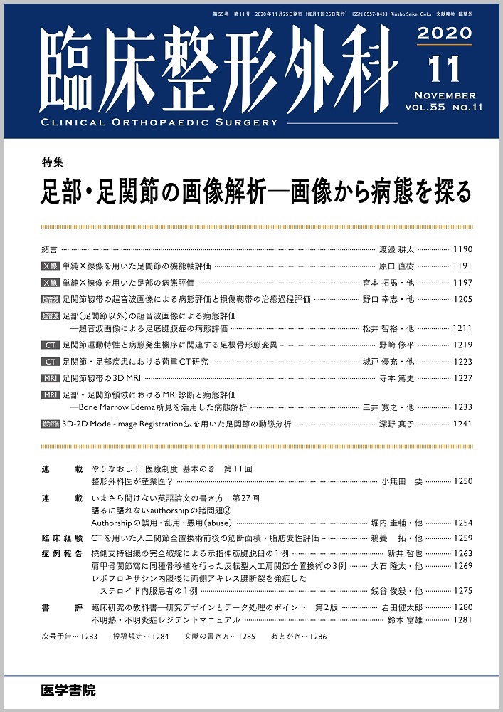
特集 足部・足関節の画像解析—画像から病態を探る
文献概要
単純X線は簡便で,侵襲も少なく,骨形態を評価できる方法であり,日常診療で最も頻繁に行われている評価方法の1つである.そのため,骨形態の時系列変化を追うのに最も適していると考えられる.本稿ではその中でも,マッピング法,横倉法,アキレス腱モーメントアームの評価法を紹介する.また,2D-3Dレジストレーションの技術は,これまで静止位でしか評価できなかったことが,動態での評価を可能とする.これにより,これまで行われていた単純X線の計測方法においても,新たな知見が得られることが期待される.
参考文献
1) Tanaka Y, Takakura Y, Sugimoto K, et al. Precise anatomic configuration changes in the first ray of the hallux valgus foot. Foot Ankle Int 2000;21(8):651-6.
2) Komeda T, Tanaka Y, Takakura Y, et al. Evaluation of the longitudinal arch of the foot with hallux valgus using a newly developed two-dimensional coordinate system. J Orthop Sci 2001;6(2):110-118.
3) 成川功一,田中康仁.外反母趾に対する水平骨切り術後における第1中足骨回内変形の矯正.J Nara Medic Asso 2009;(60)5-6:167-79.
4) 横倉誠次郎.本邦成人内外両長軸足穹窿の基準を定め扁平足の分類に及ぶ.日整会誌1928;3:331-60.
5) 米田岳史,田中康仁,藤井唯誌・他.横倉法による足縦アーチ計測点の再現性.日足会誌1999;20(2):47-51.
6) 水野祥太郎.ヒトの足.創元社.1992,p.143-155.
7) Deforth M, Zwicky L, Horn T, et al. The effect of foot type on the Achilles tendon moment arm and biomechanics. The Foot 2019;38:91-4.
8) Carbone V, Fluit R, Pellikaan P, et al. TLEM 2.0-A comprehensive musculoskeletal geometry dataset for subject-specific modeling of the lower extremity. J Biomech 2015;48(5):734-41.
9) Klein Horsman MD, Koopman HFJM, van der Helm FCT, et al:Morphological muscle and joint parameters for musculoskeletal modeling of the lower extremity. Clin Biomech 2007;22(2):239-47.
10) Rufai A, Ralphs JR, Benjamin M, et al. Structure and histopathology of the insertional region of the human Achilles tendon. J Orthop Res 1995;13(4):585-593.
11) Benjamin M, Kumai T, Milz S, et al. The skeletal attachment of tendons-tendon ‘entheses’. Comp Biochem Physio A Mol Integr Physiol 2002;133-A(4):931-45.
12) Miyamoto T, Shinohara Y, Tanaka Y, et al. Effects of achilles tendon moment arm length on insertional achilles tendinopathy. Appl Sci 2020;10(19):6631. doi:10.3390/app10196631.
13) Lenz AL, Nichols JA, Anderson AE, et al. Compensatory motion of the subtalar joint following tibiotalar arthrodesis:an in vivo dual-fluoroscopy imaging study. J Bone Joint Surg Am 2020;102(7):600-8.
掲載誌情報

