文献詳細
増刊号 ランドマークはこれだ! 局所解剖アトラス〔特別付録Web動画〕
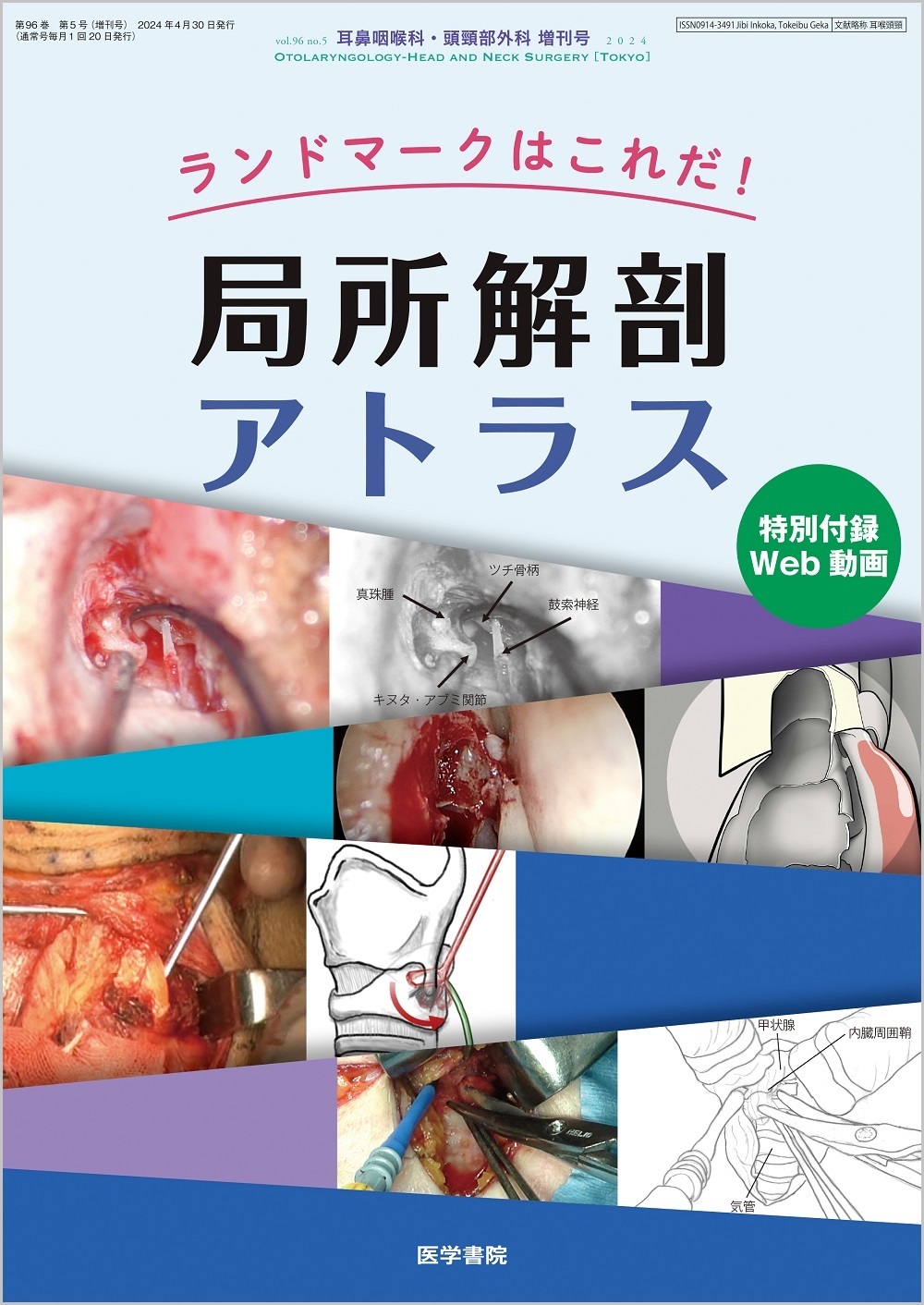
14.頭蓋底
文献概要
Point
●頭頸部外科と脳神経外科の双方の術者が,頭側から見た解剖と経鼻内視鏡下の解剖を熟知している必要がある。
●Outside-in Draf 3の際に鼻堤を削開して涙囊を露出する。涙囊後方の骨や眼窩紙様板を剝離して眼窩骨膜を同定する。
●篩骨動脈と視神経管は,眼窩紙様板を眼窩骨膜から剝離していけばおのずと見つかる。
*本論文中,動画マークのある箇所につきましては,関連する動画を見ることができます(公開期間:2029年4月)。
●頭頸部外科と脳神経外科の双方の術者が,頭側から見た解剖と経鼻内視鏡下の解剖を熟知している必要がある。
●Outside-in Draf 3の際に鼻堤を削開して涙囊を露出する。涙囊後方の骨や眼窩紙様板を剝離して眼窩骨膜を同定する。
●篩骨動脈と視神経管は,眼窩紙様板を眼窩骨膜から剝離していけばおのずと見つかる。
*本論文中,動画マークのある箇所につきましては,関連する動画を見ることができます(公開期間:2029年4月)。
参考文献
1)Castelnuovo PG, et al:Endoscopic nasal and anterior craniotomy resection for malignant nasoethmoid tumors involving the anterior skull base. Skull Base 16:15-18, 2006
2)Price JC, et al:The pericranial flap for reconstruction of anterior skull base defects. Laryngoscope 98:1159-1164, 1988
3)Chin D, et al:The outside-in approach to the modified endoscopic Lothrop procedure. Laryngoscope 122:1661-1669, 2012
4)Simmen D, et al:The surgeon's view of the anterior ethmoid artery. Clin Otolaryngol 31:187-191, 2006
5)Mundy DC, et al:A consistent endoscopic landmark to identify the anterior ethmoidal artery. Laryngoscope 134:1096-1099, 2024
掲載誌情報